Today's KNOWLEDGE Share
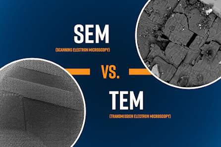
SEM Vs TEM:
To make a meaningful comparison between SEM and TEM, it’s important to note what all electron microscopes have in common. The “column” of all electron microscopes contains a series of components that are responsible for core functions.
These include:
The electron source – produces the electron beam.
Condenser lenses – directs the beam onto the sample.
Objective lens – containing the most important electromagnetic lens in the column, is responsible for forming an image of the transmitted electrons (TEM) or for forming the final focused probe that is scanned across the sample surface (SEM).
Sample chamber – holds the sample and determines the size of the sample that can be analyzed.
Detectors – collect signals to produce images.
Source:Nanoscience


No comments:
Post a Comment